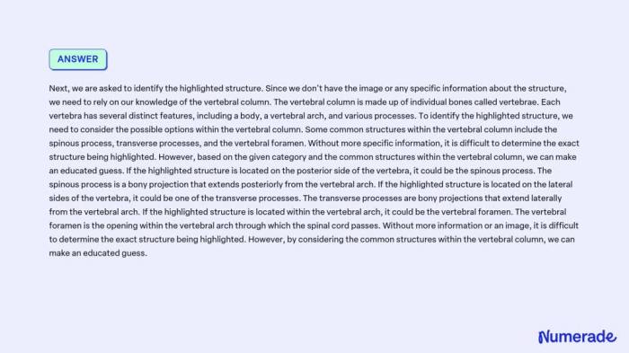
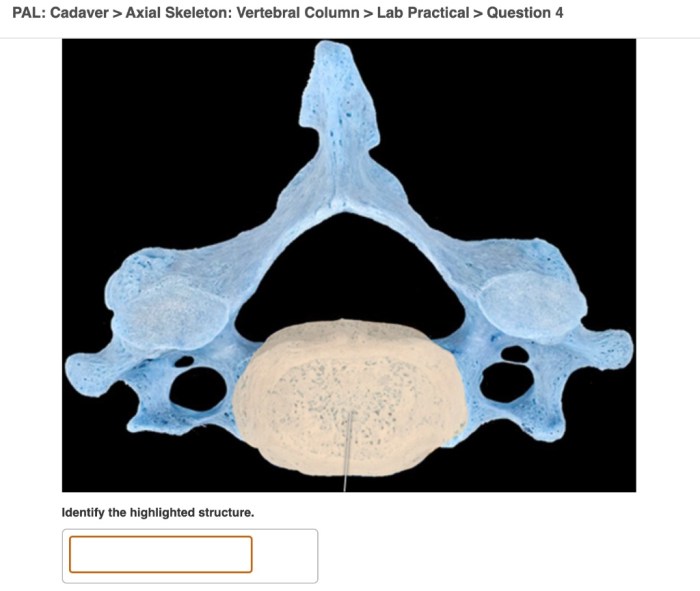
Pal cadaver axial skeleton vertebral column lab practical question 4 delves into the intricate anatomy of the vertebral column, providing a hands-on exploration of its structure and function. This practical exercise offers a unique opportunity to engage with cadaveric specimens, reinforcing theoretical knowledge and fostering a deeper understanding of this vital anatomical region.
The vertebral column, composed of a series of interconnected vertebrae, serves as the central axis of the body, supporting its weight, protecting the delicate spinal cord, and facilitating movement. Through this lab practical question, students gain firsthand experience in identifying specific vertebrae and anatomical structures, comprehending their roles in spinal stability and mobility.
Vertebral Column Overview

The vertebral column, also known as the spine or backbone, is a flexible and strong structure that forms the central axis of the human body. It consists of a series of 33 vertebrae, which are stacked one on top of the other to form a protective canal for the delicate spinal cord.
The vertebral column has several important functions, including supporting the body, protecting the spinal cord, facilitating movement, and providing attachment points for muscles and ligaments.
Regions of the Vertebral Column
- Cervical (neck): 7 vertebrae
- Thoracic (chest): 12 vertebrae
- Lumbar (lower back): 5 vertebrae
- Sacral (pelvis): 5 vertebrae, fused to form the sacrum
- Coccygeal (tailbone): 4 vertebrae, fused to form the coccyx
Pal Cadaver Axial Skeleton Lab Practical Question 4

Purpose and Objectives
The purpose of Pal Cadaver Axial Skeleton Lab Practical Question 4 is to assess students’ understanding of the anatomy of the vertebral column and its clinical significance.
The objectives of the lab practical question include:
- Identifying specific vertebrae and anatomical structures of the vertebral column
- Explaining the role of the vertebral column in supporting the body and protecting the spinal cord
- Describing common spinal disorders and injuries
Steps and Procedures
- Locate the vertebral column on the cadaver.
- Identify the different regions of the vertebral column (cervical, thoracic, lumbar, sacral, and coccygeal).
- Identify the specific vertebrae involved in Pal Cadaver Axial Skeleton Lab Practical Question 4.
- Describe the anatomical structures of the vertebrae, including the body, pedicles, laminae, spinous process, transverse processes, and articular processes.
- Explain the role of the vertebral column in supporting the body and protecting the spinal cord.
- Describe common spinal disorders and injuries, such as spinal stenosis, herniated discs, and spinal cord injuries.
Vertebral Column Anatomy

Anatomy of a Typical Vertebra
A typical vertebra consists of the following anatomical structures:
- Body: The large, cylindrical anterior portion of the vertebra that supports the weight of the body
- Pedicles: Two short, thick processes that extend posteriorly from the body and connect to the laminae
- Laminae: Two broad, flat plates that extend posteriorly from the pedicles and meet in the midline to form the vertebral arch
- Spinous process: A single, posteriorly projecting process that extends from the junction of the laminae
- Transverse processes: Two laterally projecting processes that extend from the sides of the vertebral arch
- Articular processes: Four processes that project from the superior and inferior aspects of the vertebral arch and articulate with adjacent vertebrae
Types of Vertebrae
There are five types of vertebrae, each with distinct features:
- Cervical vertebrae: Small and delicate, with a large vertebral foramen to accommodate the spinal cord
- Thoracic vertebrae: Larger and stronger, with facets on the transverse processes for articulation with ribs
- Lumbar vertebrae: The largest and strongest, with thick, robust bodies to support the weight of the upper body
- Sacral vertebrae: Five vertebrae fused to form the sacrum, which provides stability and support for the pelvis
- Coccygeal vertebrae: Four small, fused vertebrae that form the tailbone
Intervertebral Discs
Intervertebral discs are fibrocartilaginous pads that are located between adjacent vertebrae. They act as shock absorbers and allow for a limited amount of movement between the vertebrae.
Spinal Cord and Meninges
Structure and Function of the Spinal Cord
The spinal cord is a long, cylindrical bundle of nervous tissue that extends from the brainstem through the vertebral canal. It transmits sensory and motor information between the brain and the rest of the body.
Meninges
The meninges are three layers of protective membranes that surround the spinal cord:
- Dura mater: The outermost layer, a tough and fibrous membrane
- Arachnoid mater: The middle layer, a delicate and web-like membrane
- Pia mater: The innermost layer, a thin and vascular membrane that closely adheres to the surface of the spinal cord
Cerebrospinal Fluid
Cerebrospinal fluid is a clear, colorless fluid that circulates within the ventricles of the brain and the subarachnoid space surrounding the brain and spinal cord. It provides buoyancy and protection for the central nervous system.
Clinical Applications: Pal Cadaver Axial Skeleton Vertebral Column Lab Practical Question 4
Clinical Significance of the Vertebral Column and Spinal Cord, Pal cadaver axial skeleton vertebral column lab practical question 4
The vertebral column and spinal cord are vital structures that play a crucial role in maintaining mobility, supporting the body, and protecting the delicate nervous tissue within. Understanding their anatomy and function is essential for diagnosing and treating a wide range of spinal disorders and injuries.
Common Spinal Disorders and Injuries
- Spinal stenosis: A narrowing of the spinal canal that can compress the spinal cord and nerves
- Herniated discs: A condition in which the soft, inner material of an intervertebral disc protrudes through the outer layer
- Spinal cord injuries: Trauma to the spinal cord that can result in paralysis and other neurological deficits
Role of Imaging Techniques
Imaging techniques such as X-rays, CT scans, and MRIs play a crucial role in diagnosing and treating spinal conditions. These techniques allow physicians to visualize the vertebral column and spinal cord in detail, identify abnormalities, and guide treatment decisions.
Helpful Answers
What is the purpose of the vertebral column?
The vertebral column provides structural support for the body, protects the spinal cord, and facilitates movement.
What are the different regions of the vertebral column?
The vertebral column is divided into five regions: cervical, thoracic, lumbar, sacral, and coccygeal.
What are the different types of vertebrae?
There are five types of vertebrae: cervical, thoracic, lumbar, sacral, and coccygeal. Each type has distinct anatomical features.
What is the role of the intervertebral discs?
Intervertebral discs provide cushioning between vertebrae, allowing for spinal movement and absorbing shock.
What are some common spinal disorders?
Common spinal disorders include spinal stenosis, herniated discs, and spinal cord injuries.